Lysosomes are the cell’s degradation compartments and are primarily involved in the digestion of proteins, DNA, RNA, polysaccharides, and complex lipids into their respective monomers.
A well-known role of the lysosomes is to degrade and recycle the waste materials generated in their respective cells.
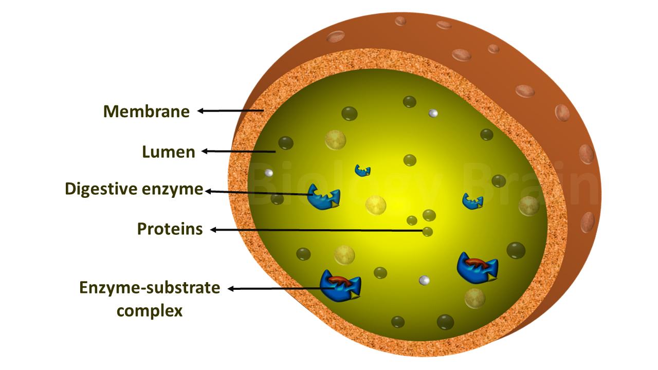
Diagram of lysosomes
- Lysosomes are formed by the budding-off of the Golgi apparatus and the collection of hydrolytic enzymes that are synthesized in the endoplasmic reticulum.
- The endoplasmic reticulum (ER) enzymes are initially tagged by mannose-6-phosphate and then they are transported to the Golgi apparatus via vesicles, where they are packed into the lysosomes.
- Structurally, lysosomes are tiny organelles, ranging from 0.1 to 1.2 µm. They have a simple structure covered by a membrane that is made up of a phospholipid bilayer.
- Inside lysosomes, various acid hydrolase enzymes are found.
- The lipids, especially the phospholipids, of all membrane systems contain a hydrophilic phosphate group and a glycerol molecule as heads and hydrophobic fatty acids as tails.
- Due to this amphipathic nature, phospholipids typically form membrane bilayers when they are kept in a solution containing water molecules. In which, the phosphate group of the lipids (hydrophilic) turns toward the hydrophilic environment (outside of the bilayer) and interacts with water molecules, while the hydrophobic fatty acid tails turn toward the inside of the bilayer (hydrophobic environment) to keep themselves away from water.
- In this way, phospholipids form cellular membranes, including cell membranes and membranes of cellular organelles such as a nucleus, mitochondria, chloroplast, lysosome, Golgi apparatus, and the endoplasmic reticulum.
Lysosome types
Lysosomes are polymorphic, which means functionally they appear in different forms.
- Primary lysosomes
- Phagolysosomes
- Autophagosomes
- Residual bodies
1. Primary lysosomes: These types of lysosomes are formed by the fusion of Golgi vesicles with late endosomes.
2. Phagolysosomes: These types of lysosomes are also called digestive vacuoles or heterophagosomes (phagosome + lysosome = phagolysosome).
3. Autophagosomes: These types of phagosomes will be formed during cellular degradation, a process called autophagy. It involves in renovation and turnover of cellular components that have previously been degraded by autophagy, eg., mitochondria, ER, peroxisomes, GC (matriphagy). Autophagy also occurs in the liver cells during starvation to provide nutrition for the remaining cellular constituents.
4. Residual bodies: These are also known as telolysosomes, which contain undigested materials.
Related topics:
References:
- Cooper GM. The Cell: A Molecular Approach. 2nd edition. Sunderland (MA): Sinauer Associates; 2000. Lysosomes. Available from: ncbi.nlm.nih.gov/books/NBK9953/
- Xu, Haoxing, and Dejian Ren. Lysosomal physiology. Annual review of physiology vol. 77 (2015): 57-80. doi:10.1146/annurev-physiol-021014-071649.
- Pu, Jing et al. “Mechanisms and functions of lysosome positioning.” Journal of cell science vol. 129,23 (2016): 4329-4339.
- Trivedi, Purvi C et al. “Lysosomal Biology and Function: Modern View of Cellular Debris Bin.” Cells vol. 9,5 1131. 4 May. 2020.
- Wilke, Sonja et al. “Crystal structure of the conserved domain of the DC lysosomal associated membrane protein: implications for the lysosomal glycocalyx.” BMC biology vol. 10 62. 19 Jul. 2012.
- Ashok K. Rout, Marie-Paule Strub, Grzegorz Piszczek, and Nico Tjandra. “Structure of transmembrane domain of lysosome-associated membrane protein type 2a (LAMP-2A) reveals key features for substrate specificity in chaperone-mediated autophagy.” The Journal of biological chemistry vol. 289,51 (2014): 35111-23.
- Eline van Meel and Judith Klumperman. “Imaging and imagination: understanding the endo-lysosomal system.” Histochemistry and cell biology vol. 129,3 (2008): 253-66.