Autophagy definition
Autophagy (“auto” means self and “phagy” means eating) is the major non-apoptotic degradation pathway in eukaryotic cells and is required for the elimination of damaged organelles, proteins, and smaller cellular debris.
It is an essential process for the cell to recycle waste and damaged elements by breaking them into smaller components. Autophagy-mediated breakdown allows cells to resynthesize newer and healthier biomolecules and organelles using degraded elements. Thus, the autophagy process can eliminate the damaged organelles and protein aggregates to reduce cellular abnormalities.
Key points of autophagy
- Involved in degradation of cytosolic metabolites and organelles including mitochondria, endoplasmic reticulum, and the nucleus by transferring them into the lysosomes.
- Autophagy is involved in the clearance of depolarized mitochondria and bulk cytosol.
- It protects homeostasis or the normal function of the body by degrading the protein aggregates in the cells.
- Involved in the turnover of broken cell organelles for new cell formation.
- Deprivation of nutrients for immune system signaling can induce the degradation of cytoplasmic material by autophagy.
- Three different classes of autophagy are Microautophagy, Chaperone-mediated autophagy (CMA), and Macroautophagy.
- The Microautophagy process uses only lysosomes for degrading the unwanted cellular components.
- Chaperone-mediated autophagy degrades substrate protein using signaling amino acid sequence (e.g. KFERQ) present in the damaged protein.
- Macroautophagy is a major autophagy process and uses phagophores to form autophagosomes, which can engulf and degrade the bulk amount of cytosolic components or organelles.
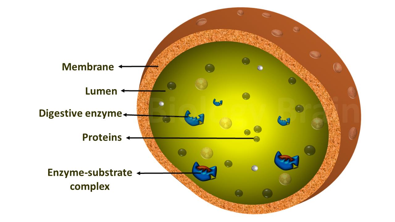
- Lysosomal enzymes contribute to the hydrolytic degradation of substrate proteins.
- Degradation of the mitochondrion is occurred by the process called Mitophagy.
- Ubiquitination also controls the multiple steps in autophagy, for instance, research studies found that a variety of ubiquitin chains are attached as selective markers on protein aggregates and dysfunctional organelles to promote the autophagy-dependent degradation.
- The process of autophagy is well organized in mammals and partially found in yeast, while completely absent in prokaryotes.
Autophagy types, and function
Stressors ranging from amino acid or nutrients deprivation to immune signaling can induce autophagy-mediated degradation. However, the elimination of particular material from the cytoplasm will occur via one of the three mentioned pathways. Elimination of damaged cellular organelles will be mediated via the macroautophagy pathway and proteins and smaller cellular debris will be eliminated via the macroautophagy pathway, while specific substrate proteins are degraded by chaperone-mediated autophagy.
1. Microautophagy
This is a nonspecific process, in which, invaginations of the lysosomal membrane itself directly engulf components of the cytoplasm.
2. Chaperone-mediated autophagy (CMA)
This is a specific or selective degradation process, in which the specialized proteins, chaperone Hsc70, and its co-chaperones will be involved. These proteins can recognize the damaged or unwanted proteins using the KFERQ amino-acid motif of the target protein. Once they recognize this sequence then they unfold the target protein. The unfolded protein binds to the lysosomal protein LAMP-2A and then translocated across the lysosomal membrane for degradation.
3. Macroautophagy
Macroautophagy is the non-selective major type of autophagy. This process is well conserved from yeast to mammals. This bulk autophagy is induced by starvation to degrade the bulk cytoplasmic components. This degradation provides the cells with essential nutrients. In the case of selective autophagy, the process occurs to clear damaged or unwanted proteins. In this autophagy process, an isolation membrane (called a phagophore) first surrounds a bulk portion of the cytoplasm or an organelle and forms an autophagosome (double membranous structure). The unwanted material contained in the autophagosome then fuses with the lysosome membrane and forms an autolysosome. In which, the hydrolytic degradation of materials takes place in autophagosomes.
4. Mitophagy (one type of Macroautophagy)
To degrade the damaged mitochondria, autophagy uses a specific type of protein receptors present on the mitochondrial membrane and recruits the core autophagic machinery to the organellar surface. The organelle is then surrounded by an isolated membrane to form a Mitophagosome which fuses with the lysosome or vacuole where the selective clearance of mitochondrion will be occurred by hydrolytic enzymes. Despite significant progress in research in recent years, some parts of the molecular machinery of mitophagy remain unclear.
Steps of the autophagy process
Autophagy can be mechanistically broken down into the following six steps
- Initiation of the isolation membrane.
- Elongation of the membrane.
- Closure of the isolation membrane
- Autophagosome formation.
- Autophagosome to lysosome fusion.
- Lysosomal degradation.
Note: These steps are similar whether invoked for the clearance of bulk cytosol or of a mitochondrion.
Autophagy inhibition by an mTOR signaling pathway
Atg 13 (Autophagy-related protein 13 Upon activation) is a very important protein for the initiation of autophagy. The activated mTORC1 phosphorylates the Atg 13. This phosphorylation prevents Atg 13 from entering the ULK1 kinase complex, which is a composite of Atg1, Atg17, and Atg101. This prevention blocks the complex structure from being recruited to the pre-autophagosomal structure present at the plasma membrane, which leads to inhibition of autophagy.
mTORC1 inhibits autophagy while at the same time induces protein synthesis and cell growth, which results in accumulations of unwanted or damaged proteins and organelles. This accumulation contributes to damage at the cellular level. Generally, activation levels of autophagy appear to decline with age. Hence, preferential activation of autophagy by scientific studies may help to promote the longevity of human life. Alterations in healthy autophagy processes have been related to cancer, cardiovascular disease, neurodegenerative diseases, and diabetes.
Data source:
Amélie Bernard and Daniel J. Klionsky. Autophagosome Formation: Tracing the Source. Dev Cell. 2 doi: [10.1016/j.devcel.2013.04.004].
Ana Maria Cuervo and Esther Wong. Chaperone-mediated autophagy: roles in disease and aging. Cell Res. 2014 Jan; 24(1): 92–104. Published online 2013 Nov 26. doi: [10.1038/cr.2013.153]
Ana Maria Cuervo. Chaperone-mediated autophagy: selectivity pays off. Trends Endocrinol Metab.
Böckler S, Westermann B.ER-mitochondria contacts as sites of mitophagosome formation. Autophagy.
G Ashrafi and T L Schwarz. The pathways of mitophagy for quality control and clearance of mitochondria. Cell Death Differ.
Orenstein SJ1, Cuervo AM. Chaperone-mediated autophagy: molecular mechanisms and physiological relevance. Semin Cell Dev Biol.
Xilouri M, Stefanis L.Chaperone mediated autophagy in aging: Starve to prosper. Ageing Res Rev. doi: 10.1016/j.arr.2016.07.001.
Yip CK, Murata K, Walz T, Sabatini DM, Kang SA. Structure of the human mTOR complex I and its implications for rapamycin inhibition. Mol Cell. doi: 10.1016/j.molcel.2010.05.017.
Zhibiao Cai, Weijun Zeng, Kai Tao, E Zhen, Bao Wang, and Qian Yang. Chaperone-mediated autophagy: roles in neuroprotection. Neurosci Bull. 2015 Aug; 31(4): 452–458. Published online. doi: [10.1007/s12264-015-1540-x]

