Structurally, the mitochondrion is a double membrane organelle that plays a central role in the production of metabolic energy in both plant and animal cells. Moreover, it’s responsible for most of the cellular energy released from carbohydrates and fatty acid catabolism. The released energy is stored in the form of ATP (adenosine triphosphate) by a process called oxidative phosphorylation.
Mitochondria Morphology
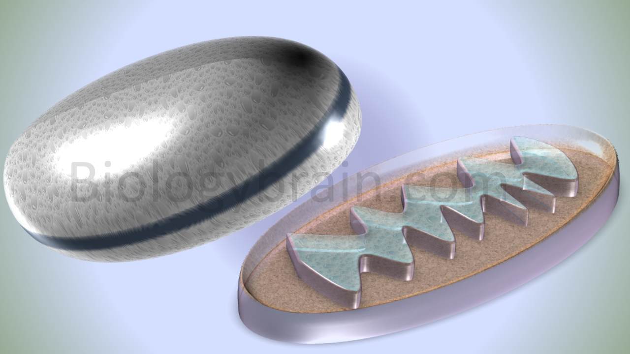
The mitochondrion is a membrane-bound organelle and it is dynamic, plastic, as well as mobile in nature. It is variable in shape, it may be spherical, filamentous, chain form, sausage-like, or cylindrical. The mitochondrion is made up of double membranes and it contains four compartments, such as outer membrane, inner membrane, inter-membrane space, and matrix.
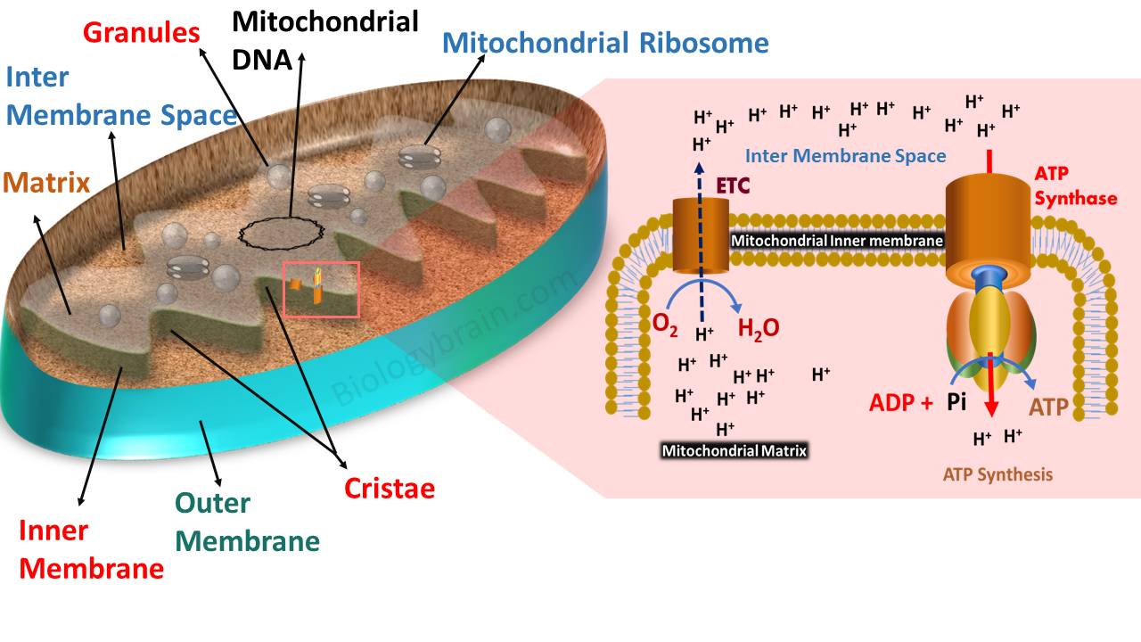
Well labelled Diagram of mitochondria and their parts
a) Mitochondrial outer membrane
Mitochondrial outer and inner membranes are closer to each other at a particular region called the contact site or contact zones.
Monoamine oxidase is an important enzyme present in the outer membrane. This enzyme has been considered a biomarker for the mitochondrial outer membrane.
The outer membrane also contains flavonoids, rotenone (pet poison), and an insensitive NADH dehydrogenase complex.
The outer membrane is permeable to several metabolites, molecules, and a few ions. However, these molecules can enter only if they are less than 5 kDa in mass.
Prions are trimeric channels found in the mitochondrial outer membrane and are involved in the permeability of molecules and ions.
Structurally, prions are composed of beta-plated sheets and are found in the outer membranes of mitochondria, chloroplasts, and the outer membranes of prokaryotic organisms, especially gram-negative bacteria.
The outer membrane also contains cholesterol, which is involved in the regulation of membrane dynamics.
b) Mitochondrial inner membrane
The mitochondrial inner membrane is more complex than other membranes as it is folded many times and separates the matrix from the intermembrane space, known as cristae. These folds are mainly formed to increase the surface area of the inner membrane.
The inner membrane has an electric potential that ranges from -160 to -120 mV. The range of electrical potential in the inner membrane is mainly due to the electrochemical proton gradient. This energy potential can be generated when the protons are shifted from the matrix (lower concentration) to the inner membrane space (higher concentration) using an electron transport system.
This membrane potential is an integral component of the proton motive force, which contributes to ATP synthesis using ATP synthase found in the inner membrane.
The inner membrane is composed of 72 to 77% proteins, 28 to 23% lipids, and 0% carbohydrates. About 20% of the membrane lipids are a composite of cardiolipin (di-phosphatidyl glycerol).
Interestingly, cardiolipin is also found on the bacterial plasma membrane. In general, cardiolipin is involved in the quaternary organization of complex-III and is also involved in the function of complex IV to promote the ATP synthase.
It is also involved in proton hopping and which contributes to ATP synthesis. It also plays a crucial role in programmed cell death.
The inner membrane contains different types of transmembrane complexes (proteins). These complexes are involved in electron transport.
NADH dehydrogenase complex (complex-I)
Succinate dehydrogenase complex (Complex-II)
Cytochrome bc complex (complex -III)
Cytochrome oxidase or terminal oxidase (complex -IV)
ATP synthase complex (complex -V)
Cytochrome oxidase is the marker enzyme for the mitochondrial inner membrane.
Cytochrome –C is the marker protein of mitochondria.
c) Mitochondrial inter-membrane space
The double membranes (outer and inner membranes) of the mitochondria create some space between them, called inter-membrane space. This space is a composition of those that are found in the cytoplasm. The proteins present in this space are distinct from the other part of the mitochondria, especially the matrix. The intermembrane space continues into the core of the mitochondria called crests or cristae.
d) Mitochondrial matrix/mitosol
A mitochondrial matrix is a liquid region that contains a complex mixture of soluble proteins and enzymes, and it is enclosed by the inner membrane. The enzymes present in the matrix are involved in the oxidation and reduction processes. The enzymes involved in the Krebs cycle are located in the matrix. These enzymes are very important to synthesize the ATP molecules, mitochondrial ribosomes, tRNAs, and mitochondrial DNA.
Mitochondrial matrix is alkaline (pH range between 7.4 to 8) in nature compared to intermembrane space (pH is 6.8 which is slightly acidic).
The matrix contains enzymes involved in the Krebs cycle and conversion of pyruvate to acetyl co.A.
The Krebs cycle enzyme, succinate dehydrogenase is found on the inner membrane and is an integral component of the electron transport system.
Malate dehydrogenase is a marker enzyme of the mitochondrial matrix.
The mitochondrial matrix contains several enzymes, which are very important in the beta-oxidation of fats.
The mitochondrial matrix contains 70s ribosomes. The ribosome in the mitochondria of a human cell is 55s type, called mitoribosomes. The 55s ribosome is divided into two subunits, such as large 25s and small 35s subunits. 25s contains 12s rRNA and the 35s subunit contains 5s rRNA and 16s rRNA.
Matrix also contains genetic material which is usually anchored to the inner membrane. Mitochondrion synthesizes most of its proteins and enzymes using its protein-synthetic machinery, hence the mitochondrion is considered a semi-autonomous organelle.
Mitochondrial dynamics
In many cells, the mitochondrion looks like a continuous form similar to the endoplasmic reticulum, hence the name mitochondrial reticulum. This form undergoes contentious organization and reorganization. The mitochondrial reticulum shows regular fission and fusion.
Fusion is mediated by mitofusin, FPZO proteins. Fission is mediated by dynamin-related proteins such as Drp-1 and Dnm-1. The morphology and distribution of the mitochondrial reticulum are very important for cellular differentiation and function. Any mutations in the fusion or fission result in the development of abnormalities. For instance, in Drosophila, a mutant prevents mitochondrial fusion leading to extensive fragmentation of the mitochondrial reticulum. This mutation blocks spermatogenesis and causes male sterility.
The degree of function of any cell mainly depends on the mass range of the mitochondria. The more mass of mitochondria is found in those cells where the energy demand is high.
Moreover, the mass of mitochondria is abundant in actively metabolizing cells, flight muscle cells of birds, cardiac muscle cells, liver cells, and sperm cells. In muscle cells, more mitochondrial mass is found near myofibrils. In flagellated cells, more mass is present beneath the basal body.
In sperm, more mass is accumulated as a sheath, which is found in the midpiece or connecting piece of the sperm. In addition, the mitochondrial mass in the cells varies with physiological conditions.
Prolonged exercise increases the mitochondrial mass in muscle cells. The scientist, GH Hogeboom was provided a piece of experimental evidence for the first time that mitochondria play a central role in cellular respiration.
References:
The Cell: A Molecular Approach. 2nd edition.Sunderland (MA): Sinauer Associates; 2000.
Friedman JR, Nunnari J. Mitochondrial form and function. Nature. 2014;505(7483):335-343. doi:10.1038/nature12985
Related topics:
| Cell organelles | Lysosome function |
| Mitochondria function | Lysosomal enzymes |
| Diagram of Lysosome | Lysosomal storage diseases |
To know more about the diagram of mitochondria and their function in the cell, keep visiting our “Biology Brain” website.