Endoplasmic Reticulum Definition
Endoplasmic reticulum (ER) is the largest single membrane-bound organelle located in the cytoplasm of all eukaryotic cells. It is a flattened network of membranous tubules and it continues with the membrane of the nucleus. The membrane-enclosed compartment of the ER is called the ER lumen. Functionally, ER membrane is very dynamic and active.
The endoplasmic reticulum interacts with the cytoskeleton and contains different forms that are majorly assigned for distinct functions.
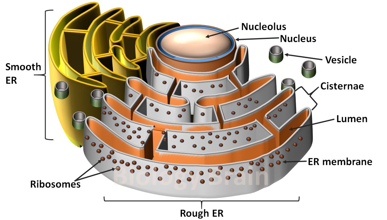
Labelled Diagram of Endoplasmic Reticulum
Types of Endoplasmic Reticulum
Two types of endoplasmic reticulum are found inside the cytoplasm depending on whether ribosomes are associated with their cytoplasmic surface of the membrane.
The ER which is devoid of ribosomes is called smooth ER (SER), while the ER that is bound with ribosomes is called rough ER (RER). Structurally, SER can be continuous with the cellular membrane called plasmalemma, while RER is continuous with the nuclear membrane. When eukaryotic cells are denatured by homogenization, the ER breaks into smaller fragments and form spherical vesicles called microsomes.
The smaller vesicles generated from RER can be identified with bound ribosomes on their surfaces and are called rough microsomes. The microsomes that are found without ribosomes on their surface are called smooth microsomes.
1. Smooth Endoplasmic Reticulum (SER)
2. Rough Endoplasmic Reticulum (RER)