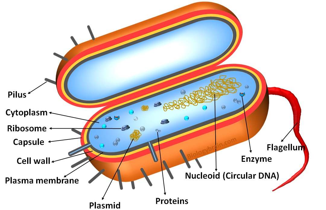
Prokaryotes are single-celled organisms belonging to the Archaea and Bacteria domains. Prokaryotic cells lack many membrane-bound cellular organelles, including lysosomes, nuclei, mitochondria, the Golgi complex, and the endoplasmic reticulum. The structure of a typical bacterial cell with its various components is depicted in the bacteria diagram provided below.
Bacteria Diagram with Labels
Bacterial cells have simpler internal structures like Pilus (plural Pili), Cytoplasm, Ribosomes, Capsule, Cell Wall, Plasma membrane, Plasmid, Nucleoid, Flagellum, etc.
Eukaryotes have been shown to be more recently evolved than prokaryotic microorganisms.
Eukaryotic cells, which make up higher organisms, evolved from prokaryotic cells. As a result, these single-celled organisms are thought to have existed on Earth four billion years ago, at the time of the earliest known life.
Both bacteria and archaea belong to the prokaryotic kingdom. The archaea includes organisms that live in some of the harshest environments, including thermophiles, methanogens, halophiles, and others. Bacteria include both harmful microbes like pathogens and helpful microbes like antibiotics, fermented goods, and essential vitamins.
Bacterial Cell Internal Structures
The internal components of bacterial cells include the Pilus (plural Pili), cytoplasm, ribosomes, capsule, cell wall, plasma membrane, plasmid, nucleoid, flagellum, etc. are more common in all prokaryotic cells.
Cell Wall
- When bacteria multiply, the cell wall, which is naturally flimsy and thin, expands significantly.
- Bacteria’s shape is controlled by the cell wall, a layer that can withstand stress.
- The majority of the bacterial cell wall is composed of murein, or peptidoglycan.
- A mixture of sugars and amino acids make up peptididoglycan.
- For the structural integrity of cell walls, peptididoglycan is crucial.
- A bacterium’s ability to survive depends on the structural integrity of the cell wall.
Bacterial Capsule
- The polysaccharide coat that most bacteria have on their cell walls is known as the bacterial capsule.
- They shield bacterial cells from the damaging effects of the environment.
- It has been discovered that capsules play a part in the colonization and infection of a number of pathogenic bacteria.
- Bacteria typically produce capsules in response to challenging conditions like higher temperatures and higher glucose concentrations.
- For instance, the production of the capsule by Aeromonas species when they enter the host blood system, an environment rich in glucose, contributes to their virulence.
- Additionally, the capsule aids in the transmission, adhesion, and survival of the bacteria as well as giving them resistance to the host immune system.
Pilus (plural Pili)
- Prokaryotic cells have tiny protein tubes that resemble thread-like structures called pili.
- They come from the plasma membrane of prokaryotic cells at the cellular level.
- Pilin, a protein, makes up the majority of the pili composition.
- Pili are in charge of the bacterial cells’ movement and are also involved in their adherence to various surfaces, which is a crucial aspect of the virulence of bacterial pathogens and aids in the spread of infection.
Plasma membrane
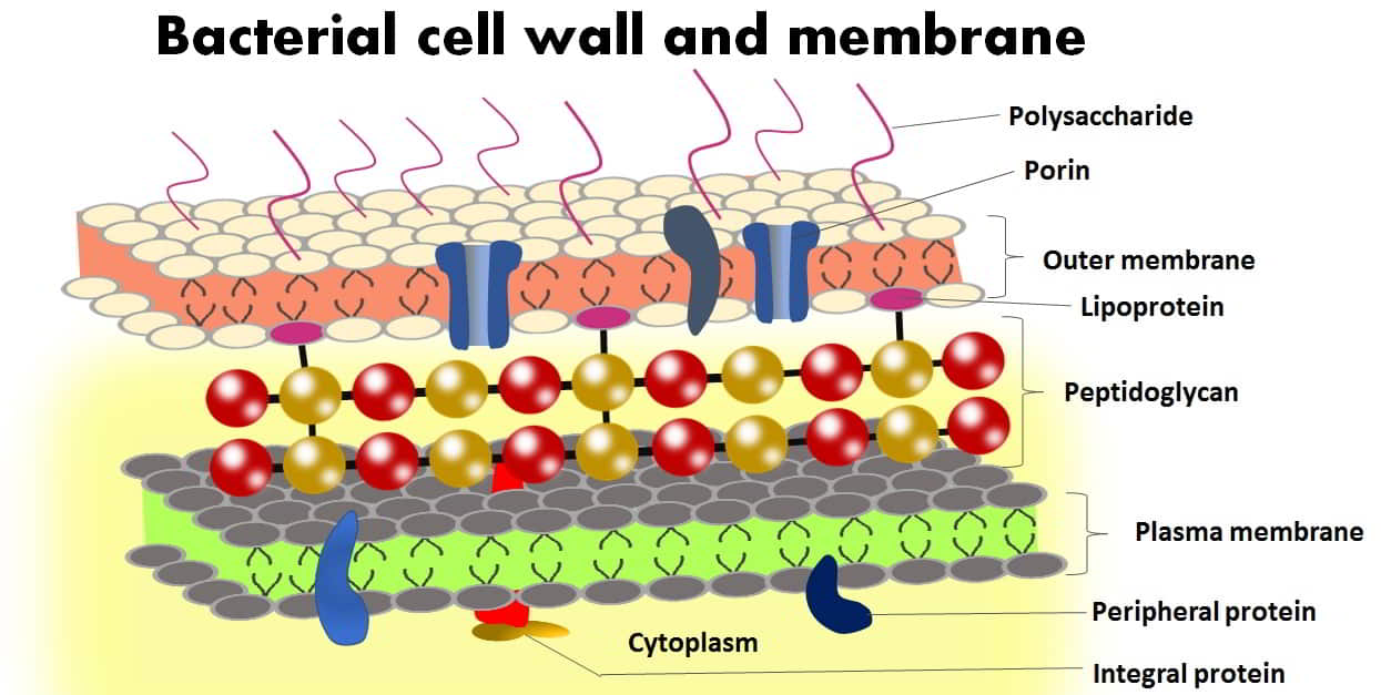
- Phospholipids, which combine to form a double-layered structure known as a phospholipid bilayer, makeup bacteria’s plasma membrane.
- The nature of the plasma membrane is amphipathic.
Hydrophobic fatty acid side chains (non-polar tails) make up the membrane’s interior while hydrophilic phosphate head groups (polar heads) make up the membrane’s exterior. - It keeps the cell’s internal environment favorable and offers general protection for the cell.
- Different proteins that are embedded in the phospholipid bilayer serve as a conduit for the selective transport of molecules, cell-to-cell communication, and virulence, among other tasks.
- The function of multi-drug resistance is also assigned to the proteins embedded in the plasma membrane.
Cytoplasm
- Organelles with membrane bindings are absent from the cytoplasm of prokaryotes.
- There is one exception, though: some prokaryotic cells also contain protein-based organelles, such as carboxysomes, as well as specialized membrane-bound organelles like magnetosomes and acidocalcisomes in their cytoplasm.
Ribosomes
- Bacteria’s ribosome (70S) is made up of two asymmetric subunits, the 50S, and the 30S subunits, which come together at a specific location on the mRNA during the start of translation.
- On the mRNA transcript, this binding site is a group of nucleotides that comes before the start codon.
- In order to synthesize bacterial proteins, each ribosomal subunit can perform a specific function.
- Ribosomes also impart a granular appearance to the cytoplasm of prokaryotes.
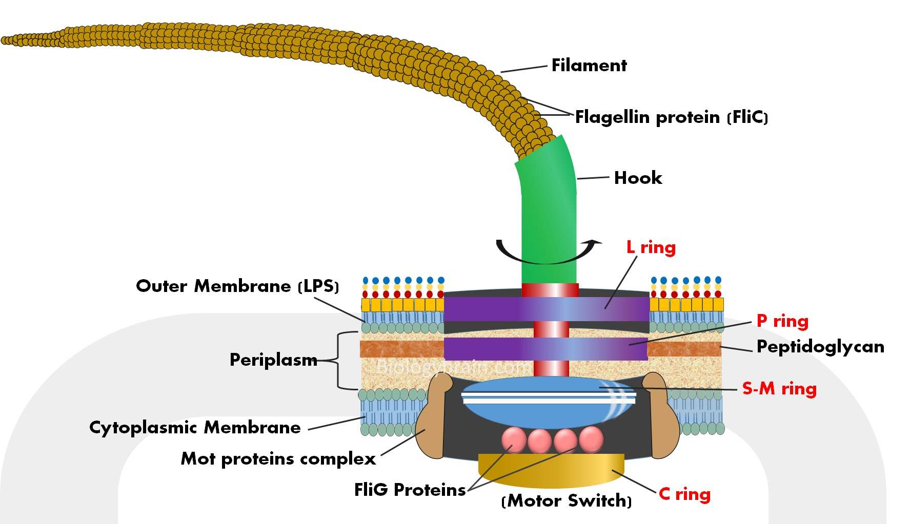
Flagellum
- Bacteria and archaea both have an organelle called the flagellum that resembles a filamentous thread.
- Movement and chemotaxis are made possible by the motile organelle known as the flagellum.
- It is made up of about 30 different types of proteins that self-assemble to create it. The copy numbers of these proteins range from a few to a few thousand.
- The ability of flagella to adhere to various environmental surfaces has also been discovered.
Nucleoid
- A nucleoid is a structure that resembles a nucleus but is not a true nucleus and has a specific location in prokaryotes.
- The prokaryotic genetic material is a tightly packed and haphazardly organized structure.
- The nuclear membrane is not present around the nucleoid.
- The majority of prokaryotic genes are found on the prokaryotic chromosome, which is circular in shape. It controls both cell division and function.
- It’s interesting to note that the prokaryotic chromosome’s length is much longer than its size.
Plasmid
- A plasmid is a tiny circular DNA molecule that coexists with the chromosomal DNA of bacteria. At the gene level, however, they are both very distinct from one another.
- Unlike bacterial nucleoids, plasmids can replicate on their own.
- In the host cell, plasmids are responsible for carrying out specific cellular tasks. Plasmids, however, generally cannot replicate without a host cell.
- The plasmid’s genes are involved in a number of processes, such as improving host survival, producing bacterial toxins, and breaking down antimicrobials.
- More than one plasmid, each with unique genes and functions, can be found in a single cell.
References
- Imada, Katsumi. “Bacterial flagellar axial structure and its construction.” Biophysical reviews vol. 10,2 (2018): 559-570. doi:10.1007/s12551-017-0378-z
- Bonnie Chaban, H Velocity Hughes, Morgan Beeby (2015). The flagellum in bacterial pathogens: For motility and a whole lot more. DOI: 10.1016/j.semcdb.2015.10.032.
- Chapter Five – Aeromonas Flagella and Colonisation Mechanisms. Advances in Microbial Physiology.
- Chapter 1 – Capsular Polysaccharides in Escherichia coli. Advances in Applied Microbiology.
- Johanna Haiko, Benita Westerlund-Wikström. The role of the bacterial flagellum in adhesion and virulence. DOI: 10.3390/biology2041242.
- Hiroyuki Terashima, Seiji Kojima, Michio Homma. Flagellar motility in bacteria structure and function of flagellar motor. Int Rev Cell Mol Biol. DOI: 10.1016/S1937-6448(08)01402-0.
- C J Jones 1 , S Aizawa (1991). The bacterial flagellum and flagellar motor: structure, assembly and function. Adv Microb Physiol. DOI: 10.1016/s0065-2911(08)60007-7.
Read Also