Flagella Definition
Flagella are thread-like or lash-like microscopic bacterial surface appendages. The term “flagellum” means whip. It can be found on the cells of both prokaryotic and eukaryotic organisms. The flagellum is mainly assigned to cellular movements (locomotion, propel, motility, or swimming) in liquid environments.
In general, three types of thread-like appendages are found on the surface of the cells, especially bacteria cells; (1) Flagella, the organelle that is responsible for locomotion, (2) Pili, the organelle that is majorly assigned to conjugation, and (3) fimbriae, the finger-like projection is involved in the attachment.
Diagram of Flagella in Bacteria
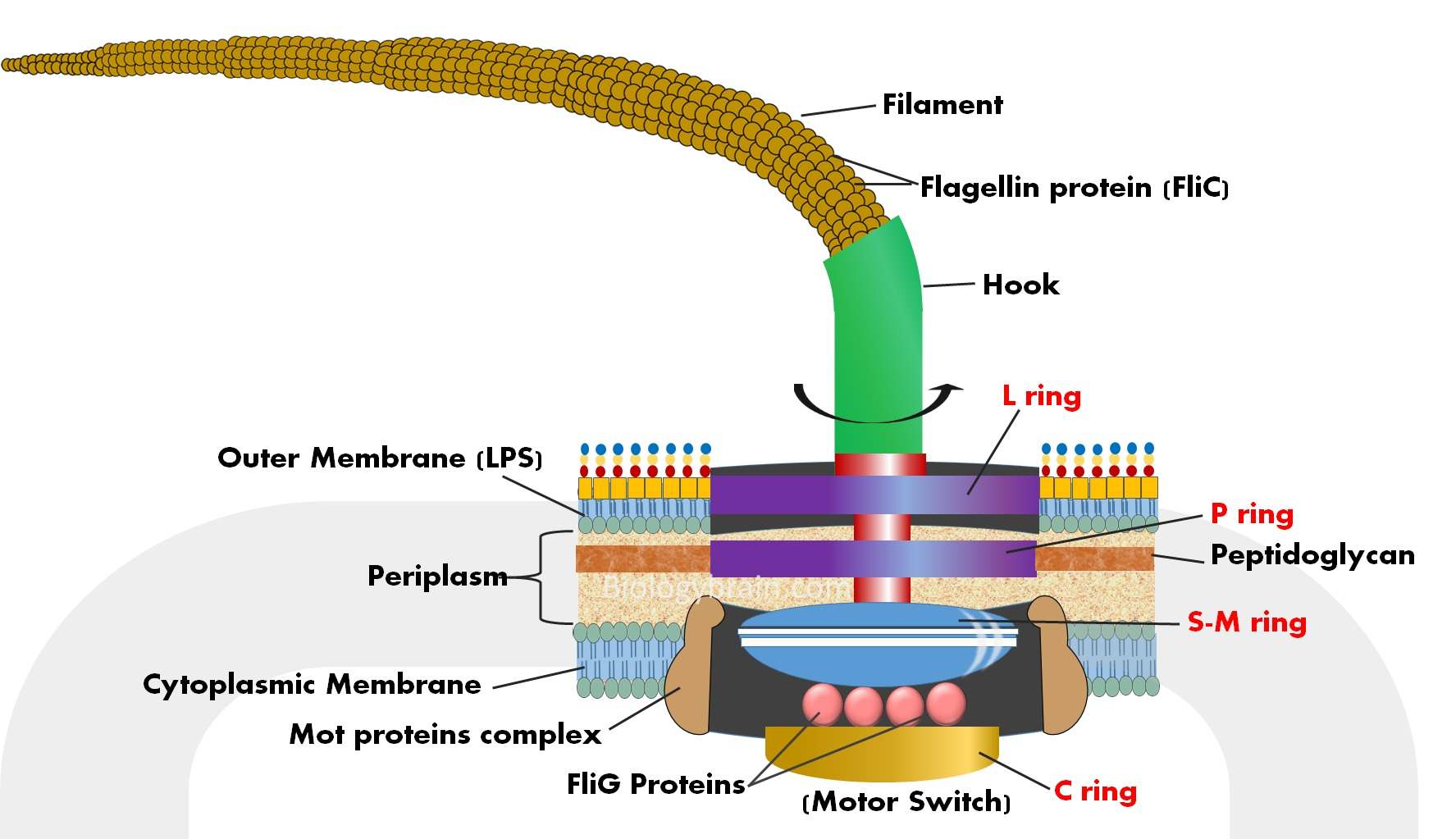
The typical flagellum of gram-negative bacteria such as E. coli is a composite of three parts.
1. The basal body of the flagella
- The basal body is a major component of the flagella of bacteria.
- It is a rod-shaped structure and a complex of different rings embedded in the cell membrane.
- To the basal body, the hook is attached and that imparts motion to the entire flagellum.
- The basal body of flagella is comprised of different rings including P ring (P for peptidoglycan), L ring (L for lipopolysaccharide), M ring (M for membrane), and S ring (S for supramembrane).
- Both M and S rings form a single complex structure called the M-S ring. The M and S rings are two different domains of the same protein family, and they perform the same function as a single unit.
- Moreover, there is another ring called the C ring (C for cytoplasmic) found beneath the inner membrane of the bacterial cells.
- FliG is a protein component of the switch complex bound to the C ring and is considered as a part of the C ring.
- Each flagellar motor switch complex is composed of about 25 FliG molecules.
- Mot proteins will combine and form an integral membrane protein complex called a motor switch.
- MotA and MotB are two different proteins that surround the M-S ring.
- These two proteins create pores in the membrane and harvest energy for mechanical rotation of flagella by proton motive force (PMF) across the cell membrane.
- Mot proteins and MS, and C rings are joined together and form a functional unit called the rotor complex, which makes a motion to the entire flagella.
2. The hook of the flagella
- There is a flexible joint between the filament and basal body called the “flagellar hook”.
- The hook is synthesized by the polymerization of 100 to 120 copies of the same protein molecule called FlgE.
- This hook is crucial for transmitting rotational torque (force) from the motor while changing its angle and that allows the flagellar filament to change its position.
3. The long filament of the flagella
- The flagellar filaments are rigid in nature. The protein flagellin is a major component of the flagellar filament.
- In the filament, the flagellin proteins are self-assembled to form a thread-like structure with a hollow core.
- In general, the length of the filament is many times the length of the bacterial cell.
- The filament ends with a cap protein (FliD).
- FliD protein controls the assembly of the filament by chaperoning and sorting flagellin proteins (FliC) after they cross the hollow filament, and leave the growing tip of the flagella.
- In the absence of capping protein, the flagellum is not synthesized, which leads to impaired locomotion of bacterial cells.
Flagella of bacteria and types
- Both gram-positive and gram-negative bacteria possess flagella on their surface.
- Structurally, flagella of bacteria are very long and filamentous.
- The number and pattern of distribution of flagella on the surface of a bacterial cells are differ from species to species.
- This unique characteristic of bacteria can be used in the identification and classification of bacterial species.
- Based on the flagellum number and arrangement, bacteria are classified into four types.
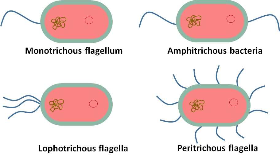
1. Monotrichous Flagellum/Monotrichous Bacteria:
It is a single flagellum, the bacterium that possesses a single flagellum is called monotrichous bacteria. Example: Vibrio cholerae.
2. Amphitrichous Flagella/ Amphitrichous Bacteria:
A single flagellum is found on each of the two opposite ends of the bacteria. This type of bacteria is called amphitrichous bacteria. Example: Campylobacter jejuni.
3. Lophotrichous Flagella/ Lophotrichous Bacteria:
The multiple flagella attached at one end of the bacteria; this is a lophotrichous bacterium. Example: Spirillium and Pseudomonas fluorescens.
4. Peritrichous Flagella/ Peritrichous Bacteria:
Multiple flagella distributed the whole surface of the bacteria. This bacterium is called a peritrichous bacterium. Example: Escherichia coli and Salmonella typhi.
The function of flagella in bacteria (flagellar movement, motility, or locomotion)
- The function of flagella in the bacteria is an energy-required process.
- Typically, the flagellar movement is rotatory in nature.
- The required energy for the flagellar motion is obtained from the proton gradient that generates proton motive force (PMF) across the cell membrane.
- No ATP has required for the function of flagella; however, the membrane-embedded proteins such as MotA and MotB form a proton channel in the membrane and generate a proton motive force for the rotation of flagella.
- The flagellar rotation can be clockwise or anticlockwise.
- When the flagellum rotates clockwise then the forward movement of bacteria ceases and cells get stuck.
- While clockwise rotation of the flagellum will pull the bacteria forward.
- In addition to the locomotor function of flagella, the flagellum is also assigned to several other functions that differ between bacteria.
- During the life cycle of bacteria: a flagellum is involved in adhesion, protein export, and biofilm formation.
References:
- Johanna Haiko and Benita Westerlund-Wikström. The Role of the Bacterial Flagellum in Adhesion and Virulence. Biology (Basel).
- S Moens, J Vanderleyden. Functions of bacterial flagella. Crit Rev Microbiol.
- Hiroyuki Terashima , Seiji Kojima, Michio Homma. Flagellar motility in bacteria structure and function of flagellar motor. Int Rev Cell Mol Biol.
- Robert M. Macnab. The Bacterial Flagellum: Reversible Rotary Propellor and Type III Export Apparatus. J bacteriol.
- Shuichi Nakamura and Tohru Minamino. “Flagella-Driven Motility of Bacteria.” Biomolecules vol. 9,7 279. 14 Jul. 2019, doi:10.3390/biom9070279.
- Tomoko Yamaguchi, Fumiaki Makino, Tomoko Miyata, Tohru Minamino, Takayuki Kato, and Keiichi Namba. “Structure of the molecular bushing of the bacterial flagellar motor.” Nature communications vol. 12,1 4469. 22 Jul. 2021, doi:10.1038/s41467-021-24715-3
- Takashi Fujii, Takayuki Kato, Koichi D Hiraoka, Tomoko Miyata, Tohru Minamino, Fabienne F V Chevance, Kelly T Hughes, Keiichi Namba (2017). Identical folds used for distinct mechanical functions of the bacterial flagellar rod and hook. DOI: 10.1038/ncomms14276.