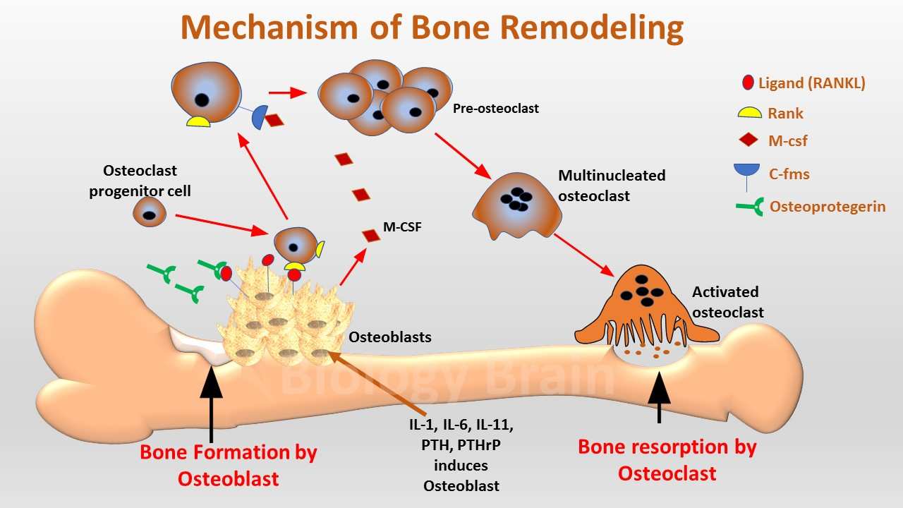
The bone remodeling process is crucial for maintaining the balance in the metabolic changes between bone formation and bone resorption.
Two specialized cells, such as osteoblast and osteoclast, are responsible for remodeling the bones.
Both bone ossification and osteoclastogenesis are mediated by the osteoblast cell, whereas osteoclast is primarily responsible for bone resorption (breakdown).
The activation of these cells ensures skeletal integrity and upholds blood mineral homeostasis throughout the lifespan of the organism. However, any changes to bone remodeling result in bone disorders.
The main factors involved in the bone remodeling process
1. Receptor activator of NFkB ligand (RANKL)
- On the membrane of osteoblast cells, the protein-ligand molecule RANKL (a cytokine of the TNF family) is expressed.
- The ligand is necessary for the development of immune cells, mammary glands, and osteoclasts.
- In the cell, RANKL can be found in two different forms, including the soluble form and the membrane-bound form.
- Both of these forms transmit signals that cause osteoclast cells to form.
- The growth and differentiation of osteoclast progenitor cells are stimulated by the binding of RANKL to its receptor, RANK (Nuclear factor-kB), during the formation of osteoclasts.
2. RANK role in the bone remodeling process
- The receptor activator of NF-κB is a receptor expressed on the membrane of osteoclast progenitor cells.
- It interacts with RANKL, and RANKL then transmits the signaling messages to intracellular transcription factors that cause osteoclasts to proliferate and differentiate.
3. Osteoprotegerin for the remodeling of bone
- Osteoprotegerin (OPG) is a cytokine receptor that belongs to the superfamily of tumor necrosis factor receptors. The TNFRSF11B gene is responsible for encoding OPG.
- OPG receptors prevent osteoclastogenesis, a process that causes excessive bone breakdown (resorption), by blocking it.
- As a result, the OPG is also known as an OCIF (osteoclastogenesis inhibitory factor).
- This inhibition process was brought about by the binding of OPG to RANKL; the OPG-RANKL complex lowers the amount of RANKL that is available to the RANK receptor.
- Therefore, the relative expressions of RANKL and OPG in bones are important regulators of the maintenance of bone mass and bone strength.
- Osteoclast activity is decreased by PGP concentrations greater than RANKL concentrations, which slows down bone remodeling.
4. Macrophage colony-stimulating factor (MCSF) and its function in the remodeling of bone
- MCSF is an essential component for the bone to remodel.
- MCSF plays a role in tissue macrophages as well as osteoclast and monocyte proliferation and differentiation.
- Additionally, lysosome synthesis, lysosome function, and the development of the ruffled border in osteoclast cells are all significantly influenced by MCSF.
- Ruffled borders are essential for increasing the surface area of osteoclast cells, which makes it easier for digestive enzymes to be delivered and for an acidic environment to be created, which enables osteoclasts to break down bone.
- It has been discovered that osteoclast cells’ lysosomes play a crucial role in the digestion of ingested bone fragments.
- A lack of MCSF causes abnormal bone formation in the bone tissue.
5. c-Fms (M-CSF receptor) and its function in the remodeling of bone
- A receptor called c-Fms is present on the membrane of osteoclast progenitor cells, and it enables M-CSF ligand to bind to tissue macrophages and promote the growth of osteoclasts and monocytes.
6. Interleukins and their role in the remodeling of bone
- Cytokines, which include interleukins, play a crucial role as messengers in cellular signaling.
- When bone is being remodeled, T-cells secrete interleukins to stimulate osteoblast cells, which then release the signaling molecule to promote osteoclast proliferation and differentiation.
7. Serine-threonine protein kinase Akt role in the remodeling of bone
- Under the control of numerous external and internal factors, both bone mass and its turnover are maintained with an equilibrium between bone formation and bone resorption.
- One of the key players in the signaling process mediated by bone anabolic factors is phosphoinositide-dependent serine-threonine protein kinase or Akt.
- Low turnover of osteopenia is caused by bone cell dysfunction and lower levels of Akt expression in osteoblasts and osteoclasts.
- The proliferation, differentiation, and survival of osteoblasts and osteoclasts are essential for maintaining bone mass and turnover, and Akt has been identified as a key modulator in this process.
Research on RANKL
- In previous studies, scientists from the University of Arkansas for Medical Sciences, USA, performed experiments on RANKL (membrane-bound and soluble form) and stated that RANKL is an important factor in osteoclast formation.
- For their studies, they generated mice, which have a sheddase-resistant enzyme (a protease enzyme that shows potent RANKL sheddase activity) for RANKL release.
- Thus, this mouse lacks a soluble RANKL in its blood circulation.
- From their experiments on mice, they found that deficiency of soluble RANKL affects the bone structure or its mass in mice but reduces osteoclast cell number and increases cancellous bone mass in adult mice.
- They stated that the bone loss caused by estrogen deficiency, however, is unaffected by the absence of the soluble form of RANKL.
- While, the lymphocyte number, lymph node development, and mammary gland development are also unaffected by the lack of soluble RANKL.
- Finally, they concluded that the membrane-bound form of RANKL is sufficient for most functions of this protein, but the soluble form of RANKL is essential for the physiological process of bone remodeling in adult mice.
References
- Brendan F. Boyce, M.D. and Lianping Xing, M.D., Ph.D. Functions of RANKL/RANK/OPG in bone modeling and bone remodelling. Arch Biochem Biophys. 2008 May 15; 473(2): 139–146.
- Sims NA1, Jenkins BJ, Nakamura A, Quinn JM, Li R, Gillespie MT, Ernst M, Robb L, Martin TJ. Interleukin-11 receptor signaling is required for normal bone remodeling. J Bone Miner Res. 2005 Jul;20(7):1093-102.
- Bozec A1, Zaiss MM. T Regulatory Cells in Bone Remodelling. Curr Osteoporos Rep. 2017 Jun;15(3):121-125.
- Mark C.HorowitzPhD. The Role of Cytokines in Bone Remodelling. Journal of Clinical Densitometry.
- Soluble RANKL contributes to osteoclast formation in adult mice but not ovariectomy-induced bone loss. Nature Communications volume 9, Article number: 2909 (2018).