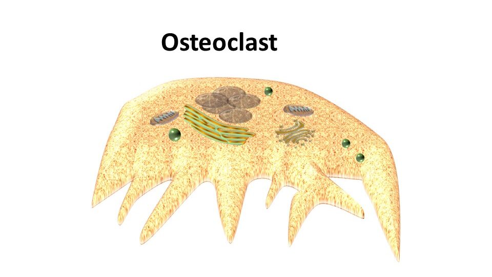
Osteoclast Definition
An osteoclast (plural “osteoclasts”) is a multinucleated cell found only in bones. It plays a crucial role in bone absorption and regeneration. Bone is a dynamic connective tissue in vertebrates. An osteoclast digests bulk molecules such as hydrated proteins and minerals into small elements using enzymes, collagenase and acid phosphatase, and some potent acids; a process called bone resorption.
Key Points of Osteoclast
- Osteoclast was discovered by Albert Kolliker in 1873 and he proposed that osteoclast function is mainly designed to digest the bone.
- The term “osteoclast” is used in bone-cell biology, derived from Greek (osteo means “bone” and clast means “broken”).
- In vertebrates, bone development and bone resorption are closely coupled mechanisms that are responsible for bone remodeling.
- The osteoclast is differentiated (or derived) from hemopoietic progenitor cells of the monocyte-macrophage
- It contains a large number of cellular organelles such as mitochondria, lysosomes, and vacuoles.
- The vacuole is the very essential organelle in osteoclast cells to digest the engulfed bone fragments.
- Osteoblasts are other types of bone cells involved in osteoclastogenesis through cell-to-cell contact with osteoclast progenitors.
- Osteogenesis imperfecta is a brittle bone disease caused due to genetic mutations, especially the genes, COL1A1, and COL1A2 which are responsible for bone formation.
- Osteoporosis is another process, in which the rate of bone formation (osteogenesis) is lower than bone resorption. The resulting condition is reduced bone mass.
- The deficiency of estrogen enhances the activity of osteoclasts, which results in the enhancement of osteoclast formation and bone resorption.
- In women, after menopause, the activity of osteoclasts is increased due to the low level of estrogen at this point.
- Any alterations in testosterone levels in men may also increase the activity of osteoblast cells.
- The fluctuations in sex hormone levels in both men and women lead to emerging bone problems, including bone fractures or trauma.
- The combination of estrogen and progesterone reduces absorption and increases bone strength.
- Odontoclast is another type of cell that also shows a similar function in human deciduous teeth.
Where is osteoclast present and what are its main cellular characters?
As mentioned above, osteoclast is a bone cell and is present in pits of the bone surface, which are called resorption bays or Howship’s lacunae. It contains homogeneous cytoplasm with a foamy appearance. This foamy character of the cytoplasm is due to the presence of more amount of vesicles and vacuoles. The vacuoles such as lysosomes are filled with the enzyme, acid phosphatase. The acid nature of osteoblast cells allows researchers to identify these cells through the staining method. The staining method also gives information on how much amount of tartrate-resistant acid phosphatase (TRAP) and cathepsin K are present in the osteoclast cells. Moreover, osteoclasts contain more amount of thin rough endoplasmic reticulum and an extensive Golgi complex.
The function of osteoclast
Osteoclasts can synthesize various enzymes, e.g. acid phosphatase, that dissolves organic proteins, especially collagen as well as inorganic phosphorus and calcium of the bones. Mineralized bone is first broken down into small fragments then the osteoclasts engulf these fragments and send them to vacuoles. Then further digestion takes place by the enzymes present in the vacuoles. Calcium and phosphorus produced by the breakdown of the mineralized bone are released into the bloodstream. Unmineralized bone (osteoid) is protected from osteoclastic resorption.
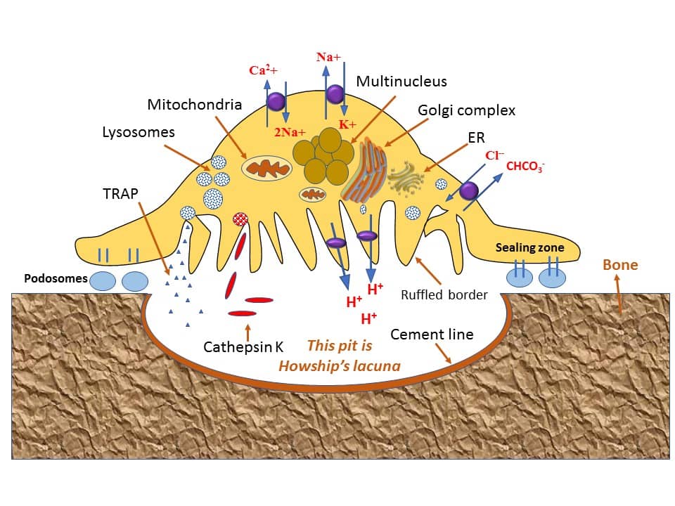
Cathepsins are proteolytic enzymes involved in the breakdown of collagen and other noncollagenous proteins. Especially, the cathepsin K enzyme is produced by osteoclasts and is involved in bone resorption by degrading the bone structural proteins. Any mutations in the collagen gene lead to pycnodysostosis, a hereditary osteopetrosis disease, which is a result of a deficiency in the functional cathepsin K.
In acidic conditions, Cathepsin K will show its optimal enzymatic activity. Initially, cathepsin is synthesized as a proenzyme with a molecular weight of 37kDa, and later it is activated by autocatalytic cleavage and generates a ~27kDa weight active fragment. Once the active form is generated then it will be involved in the proteolytic process.
Upon polarization of the osteoclasts at the site of bone resorption, cathepsin K is released into the resorptive pit from the ruffled border. Cathepsin K transmigrates across the ruffled border by intercellular vesicles and is then released by the functional secretory domain. Within these intercellular vesicles, cathepsin K along with reactive oxygen species will be generated by TRAP and they degrade bone extracellular matrix.
What are the metabolites that regulate the function of osteoclast?
Several hormones are found to regulate osteoclast function, especially the hormones secreted by the thyroid glands such as parathyroid hormone (PTH) (from the parathyroid gland) and calcitonin (from the thyroid gland), and a growth factor such as interleukin 6 (IL-6). IL-6 is one of the main factors in osteoporosis disease (an imbalance condition between bone resorption and bone formation). While osteoclast function is also regulated by the interaction of two molecules released from osteoblasts, namely osteoprotegerin and RANK ligand. The fact these molecules also regulate osteoclast differentiation.
Osteoclast differentiation
Differentiation of osteoclasts occurs in two ways, which are mainly defined based on osteoblast involvement.
Osteoblast dependent osteoclast differentiation
Recent studies on interactions between Receptor Activator of NF-Kappa B (RANK) and RANK ligand (RANKL) discovered that osteoblasts play an important role in osteoclast cell differentiation. Osteoblasts express a membrane-associated factor RANKL on their cell membranes. While, osteoclast precursor cells express a receptor, RANK on their membranes. RANKL and RANK interactions (cell-cell interaction) can recognize and differentiate osteoclast precursor cells into osteoclasts. Hence, osteoblast is also responsible for osteoclast differentiation.
Osteoblast-independent differentiation of Osteoclast
Also, recent studies have found that the lipopolysaccharide and inflammatory cytokines such as tumor necrosis factor receptor-alpha and interleukin-I directly induce osteoclast differentiation and function. This mechanism is independent of the RANKL-RANK interaction. Interferon-gamma and transforming growth factor-beta superfamily members are also found to be important factors for osteoclastogenesis. These studies have provided new strategies for analyzing the molecular mechanisms involved in osteoblast and osteoclast differentiation.
Frequently asked questions:
1. Which of the following is associated with osteoclast resorption of bone?
A. Osteoclast decreases calcium levels in the blood
B. Osteoclast inhibits bone resorption
C. Osteoclast secrets digestive enzymes for bone resorption
D. It activates the parathyroid gland if calcium levels are high in the blood
Answer: C
2. What is osteoclast?
Answer: Osteoclast is a large multi-nucleated cell that secretes digestive enzymes like collagenase and acid phosphatase, and releases some important acids.
3. Which hormone increases osteoclast activity to release more calcium ions into the bloodstream?
A. Thyroxine
B. Parathyroid hormone
C. Progesterone
D. Estrogen
Answer: B (Low levels of calcium in the blood stimulates the parathyroid gland to release parathyroid hormone (PTH), which activates osteoclasts to release calcium from the bones)
Data source
- Enomoto H, Shiojiri S, Hoshi K, Furuichi T, Fukuyama R, Yoshida CA, Kanatani N, Nakamura R, Mizuno A, Zanma A, Yano K, Yasuda H, Higashio K, Takada K, Komori T. Induction of osteoclast differentiation by Runx2 through receptor activator of nuclear factor-kappa B ligand (RANKL) and osteoprotegerin regulation and partial rescue of osteoclastogenesis in Runx2-/- mice by RANKL transgene. J Biol Chem.
- Väänänen HK, Härkönen PL. Estrogen and bone metabolism. Maturitas.
- Sunyer T, Lewis J, Collin-Osdoby P, Osdoby P. Estrogen’s bone-protective effects may involve differential IL-1 receptor regulation in human osteoclast-like cells. J Clin Invest.
- Katagiri T and Takahashi N.Regulatory mechanisms of osteoblast and osteoclast differentiation. Oral Dis.
- Udagawa N. The mechanism of osteoclast differentiation from macrophages: possible roles of T lymphocytes in osteoclastogenesis. Bone Miner.
- Sasaki T. Differentiation and functions of osteoclasts and odontoclasts in mineralized tissue resorption.Microsc Res Tech.
- Wang Z, McCauley LK. Osteoclasts and odontoclasts: signaling pathways to development and disease. Oral Dis.